|
|
|
Milosavljevic
P.D. *, Milenkovic M. **, Colic M. *, Dimitrijevic
M.**.
*Institute of Medical Research, Military Medical Academy,
Crnotravska 17, 11002 Belgrade and **Department of Microbiology and Immunology,
Faculty of Pharmacy, Belgrade, Yugoslavia
Introduction, Material & Methods
An experimental autoimmune myocarditis in genetically
susceptible DA rats has been developed with the aim to investigate immunohistological
changes in the course of the disease with special reference to the expression
of the dendritic cell (DC) markers.
Male DA rats were immunized with porcine cardiac myosin
in complete Freunds adjuvant (CFA). Animals were sacrificed at the different
time points after the disease induction. Immunopathological changes of
the heart tissue and phenotype of infiltrating cells were examined using
immunoperoxidase and immunoalkaline phosphatase methods and monoclonal
antibodies specific for rat MHC class II (OX6), CD80 and CD86 (costimulatory
molecules) and OX62 (rat dendritic cells).
Results
In the hearts of control animals DC, predominantly localized
in the interstitium, were OX6hi CD86lo CD80- OX62-. In the hearts of diseased
animals the first changes were observed after one week, which were manifested
as an increase in the expression of MHC class II (OX6) molecules. Rare
perivascular DC, phenotypically OX6+ OX62+ CD86+ CD80lo appeared. On day
16, large cell infiltrates were observed. DC in the infiltrates were numerous
and expressed strongly OX6, OX62 and CD86 markers, whereas the expression
of CD80 was still lower. The number of OX62+ DC was lower then the numbers
of other cells suggesting that CD80, CD86 as well as MHC class II molecules
were also present on other cell types in the infiltrates, probably on macrophages.
The number of DC in the interstitium was not significantly changed. Thirty
two days after the disease induction, inflammatory infiltrates were reduced
followed by the decrease in the DC number.
 .
.
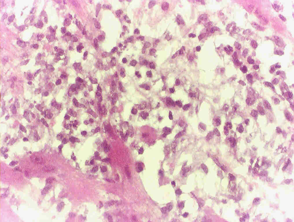
Inflammatory infiltrates in heart muscle
with massive
destruction of heart tissue 16 days after
immunization
Conclusion
Our immunohistochemical analysis showed that ED1+
MØ and DC constitute a most significant populations of cells
in inflammatory infiltrates which probably migrated from circulation rather
than from the heart interstitium.
If you have comments or suggestions, email me at
petarm@EUnet.yu